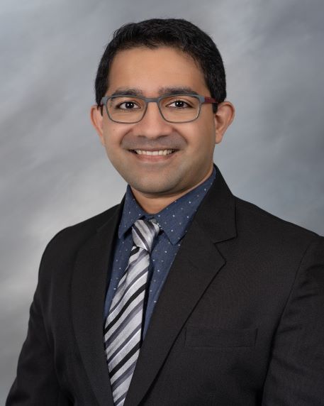
| Faculty Profile |
|
Office Address
Wayne State University School of Medicine
Department of Neurology, Suite 8D-UHC
4201 St. Antoine
Detroit, MI 48201
Phone: (313) 577-1245
Fax: (313) 745-4216
Narrative Bio
Sagar Buch, PhD is an Assistant Professor (Research) with the Department of Neurology here at Wayne State University, School of Medicine. Dr. Buch has more than 10 years of experience in the field of neuroimaging and implementing advanced magnetic resonance imaging (MRI) quantitative techniques. Dr. Buch’s background and expertise has been in MRI physics, development and optimization of MR-based protocols for quantitative imaging such as quantitative susceptibility mapping (QSM). He is experienced in MR angiography/venography, susceptibility weighted imaging (SWI) and signal processing for studying the cerebral microvasculature with an eye toward their effects in normal aging, as well as in neurodegenerative and neurovascular diseases.
QSM utilizes the MRI phase images that exhibit the perturbation of the static magnetic field of the MRI scanner across the different biological tissues. This perturbation is dependent on a tissue’s magnetizability (or: magnetic susceptibility). QSM measures these perturbations and computes the underlying spatial distribution of susceptibility by solving physics-based inverse mathematical problem, a process to which he has made several contributions including the development of a 3D numerical model of the whole brain that has been used to analyze the effects each individual step involved in the QSM process and in developing a technique to map structures that exhibit no discernable MRI signal such as the sinuses and teeth. Lately, his focus is on using an optimized high-resolution protocol for ultrasmall superparamagnetic iron oxides (USPIO)-based imaging. This study involves studying the vascular pathophysiology, implementing image processing and reconstruction to plan the next generation of microvascular imaging, which is referred to as MICRO or, Microvascular In-vivo Contrast Revealed Origins to study microvascular details in animals and in humans. This in vivo protocol allows us to reveal the underlying vascular details in healthy subjects to study the effects of normal aging on the vasculature; and vascular abnormalities such as submillimeter-sized lesion-centric developmental venous anomalies (DVA) that was only thought to be possible on the cadaver brain data. This project has sparked new-found interest in the field of neurovascular imaging.
Dr. Buch received his MASc in 2012 and his PhD in 2015 from McMaster University in Canada. He was a MR Research Consultant, with the Department of Radiology, Wayne State University from 2019 – 2022. He worked as a MR Research Scientist, with the MRI Institute for Biomedical Research, Waterloo, Ontario, Canada from 2015–2017. He also held the position of Assistant Biomedical Engineer at S.L. Raheja Hospital and Diabetic Research Center in, Mumbai, India in 2009.
Dr. Buch was awarded the Potchen–Passariello Award from the Society for Magnetic Resonance Angiography (SMRA) In 2021, and a Summa Cum Laude Award, from the International Society for Magnetic Resonance in Medicine (ISMRM) in 2020.
Department URL
https://neurology.med.wayne.edu/
Academic Rank
Assistant Professor (Research)
Department of Neurology
Undergraduate
Bachelor of Engineering (BE) University of Mumbai, India 2010
Major: Biomedical Engineering.
Graduate
Doctor of Philosophy (PhD) (Biomedical Engineering), McMaster University, Canada 2015
Master of Applied Science (MASc) (Biomedical Engineering), McMaster University, Canada 2012
Suffix
PhD
Specialties
Magnetic Resonance Imaging (MRI)
Publications
Research Description
We are also collecting several PD patients' MICRO data from a different site (London, ON, Canada). So, if you are interested in vascular research for MS and/or PD, please feel free to contact me. Thank you!
